
TL;DR

Prime made for students and young adults
- Fast, free delivery for dorm and study essentials
- Prime Video and Amazon Music included
- Member-only deals
Indian scientists have produced the most detailed 3D atlas of the human brainstem, a breakthrough that could improve understanding of brain functions and neurological disorders. The development is confirmed and marks a significant advance in neuroscience tools.
Indian scientists have announced the development of the most detailed 3D atlas of the human brainstem, a comprehensive mapping project that enhances understanding of this critical brain region. This breakthrough, confirmed by the research team, represents a significant advancement in neuroanatomical mapping and could impact future diagnostics and treatments for neurological disorders.
The project was led by a team at the Indian Institute of Science, Bangalore, who utilized advanced imaging techniques and computational modeling to produce a high-resolution, three-dimensional map of the human brainstem. The atlas captures intricate neural structures with unprecedented detail, surpassing previous models in resolution and accuracy.
According to Dr. Anjali Rao, the lead researcher, the atlas integrates data from post-mortem brain samples using high-field MRI scans combined with histological analysis. The team states that this comprehensive approach allows for precise identification of nuclei, fiber tracts, and vascular structures within the brainstem, which is vital for understanding its role in vital functions like breathing, heartbeat regulation, and consciousness.
While the project is still in the validation phase, the team has shared preliminary findings in a peer-reviewed publication, confirming the creation of this detailed 3D model. The researchers emphasize that this atlas could serve as a foundational tool for neuroscientists, neurologists, and neurosurgeons worldwide.
Implications for Neuroscience and Medical Research
This development provides a detailed representation of the brainstem, which is a critical area of the brain involved in essential functions such as respiration, heart rate, and consciousness. A comprehensive 3D atlas can support improved understanding of its structure, potentially aiding in diagnosis and treatment planning for conditions like stroke, Parkinson’s disease, and brainstem tumors.
This atlas may also facilitate further research into neural pathways and disease mechanisms, contributing to the development of targeted therapies. The project demonstrates India’s ongoing efforts in neuroimaging and computational neuroscience, aligning with global research initiatives in this field.
As an affiliate, we earn on qualifying purchases.
Advances in Brain Mapping and Indian Neuroscience
Over the past decade, brain mapping has seen significant progress through the use of high-resolution imaging and computational methods. Previously, detailed models of the brainstem were limited by the resolution of available data. While international efforts have produced some 3D models, this recent Indian project offers a higher level of detail.
India has increased investment in neuroscience research, with institutions like the Indian Institute of Science and the All India Institute of Medical Sciences leading various projects. This new atlas builds on earlier neuroimaging studies and reflects ongoing efforts to develop indigenous tools for brain research.
The development aligns with global initiatives aimed at understanding brain structure-function relationships, especially in relation to neurological diseases. The Indian team’s work contributes to filling a gap in neuroanatomical resources with a comprehensive model.
“This 3D atlas provides a detailed view of the brainstem, which can support further research into its anatomy and functions.”
— Dr. Anjali Rao, Lead Researcher
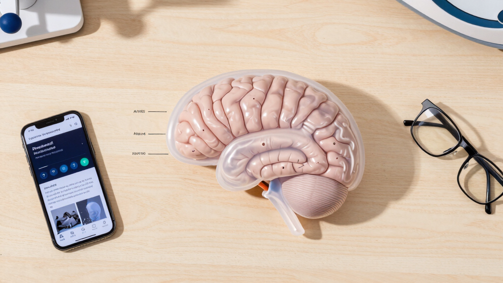
neuroscience 3D brain atlas models
As an affiliate, we earn on qualifying purchases.
As an affiliate, we earn on qualifying purchases.
Validation and Clinical Application Uncertainties
While the team has published preliminary findings confirming the creation of the atlas, it remains to be seen how widely it will be adopted or integrated into clinical practice. The validation process is ongoing, and additional peer review and testing are necessary to establish its accuracy and usefulness in medical settings.
The extent to which this atlas will be used in surgical planning or diagnostics depends on further validation and integration with existing medical imaging tools.
As an affiliate, we earn on qualifying purchases.
Next Steps for Validation and Global Collaboration
The Indian team plans to publish detailed validation studies in peer-reviewed journals over the coming months. They also intend to collaborate with international neuroscience and medical institutions to evaluate the atlas’s utility in clinical diagnostics and surgical planning.
Future research will focus on integrating this atlas with functional imaging data to better understand brainstem activity and its role in neurological diseases. Efforts are also underway to develop accessible software tools for use by clinicians and researchers worldwide.
As an affiliate, we earn on qualifying purchases.
Key Questions
How does this new brainstem atlas differ from previous models?
The new atlas offers higher resolution and more detailed visualization of neural structures, nuclei, and fiber tracts compared to earlier models, supporting more precise neuroanatomical analysis.
What potential medical benefits can this atlas provide?
It may assist in diagnosis, surgical planning, and treatment of brainstem-related neurological conditions, and support research into brain functions and disease mechanisms.
Is this atlas available for clinical use now?
No, it is currently in the validation stage. Additional testing and peer review are needed before it can be adopted in clinical practice.
What techniques were used to create this atlas?
The team utilized high-field MRI scans combined with histological analysis of post-mortem brain samples to develop the detailed 3D model.
How does this development position India in neuroscience research?
This project highlights India’s growing capabilities in advanced neuroimaging and computational neuroscience, contributing to global efforts in brain research.
Source: hn
Halloween Picks
halloween
As an affiliate, we earn on qualifying purchases.